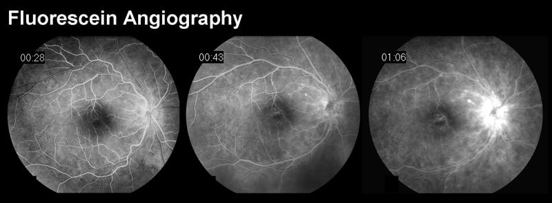
FA requires the use of a dedicated fundus camera equipped with excitation and barrier filters. Fluorescein dye is injected intravenously, usually through an antecubital vein with sufficient speed to produce high contrast images of the early phases of the angiogram. White light from a flash is passed through a blue excitation filter. Blue light (wavelength 465-490 nm) is then absorbed by unbound fluorescein molecules, and the molecules fluoresce, emitting light with a longer wavelength in the yellow-green spectrum (520-530nm). A barrier filter blocks any reflected light so that the images capture only light emitted from the fluorescein. Images are acquired immediately after injection and continue for ten minutes depending on the pathology being imaged. The images are recorded digitally or on 35mm film.

You’ll see that when it’s time to make an appointment with a specialist in Retina and Vitreous diseases, you can trust your eye health to the team at Inland Valley Retina Medical Associates, Inc.